
When evaluating neurological conditions, two of the most commonly used diagnostic tools are MRI and EEG. Both technologies provide valuable information about the brain, but they work in very different ways. Understanding how MRI and EEG differ—and why combining them can improve diagnostic accuracy—is essential for patients seeking advanced neurological evaluations.
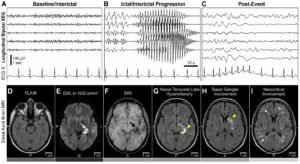
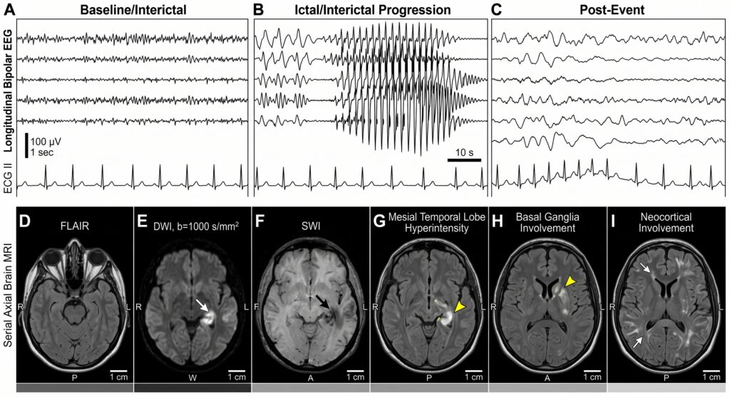
MRI focuses on the brain’s physical structure, while EEG measures electrical activity and brain function in real time. Individually, each test offers important insights. However, when combined through advanced neuroscience software imagery, clinicians can achieve a far more comprehensive understanding of neurological health.
At Applied Neuroscience INC. in St. Petersburg, Florida, integrated clinical neuroscience scans combine advanced imaging technologies to support more accurate evaluations for ADHD, traumatic brain injuries, epilepsy, cognitive disorders, and other neurological conditions.
What Is an MRI?
MRI stands for Magnetic Resonance Imaging.
MRI uses:
- Powerful magnets
- Radio waves
- Computer-generated imaging
to create detailed pictures of the brain’s anatomy.
MRI scans are commonly used to identify:
- Brain tumors
- Structural abnormalities
- Stroke damage
- Inflammation
- Brain injuries
- Degenerative neurological conditions
MRI is considered one of the most effective tools for visualizing physical changes in the brain.
What Is an EEG?
EEG stands for Electroencephalography.
EEG measures electrical activity generated by brain cells using sensors placed on the scalp.
Unlike MRI, EEG does not produce detailed anatomical images. Instead, it records:
- Brainwave activity
- Functional neurological patterns
- Electrical communication
- Real-time neural activity
EEG is commonly used to evaluate:
- ADHD
- Epilepsy
- Sleep disorders
- Cognitive dysfunction
- Seizure activity
- Neurological abnormalities
EEG provides information about how the brain functions rather than how it looks structurally.
MRI vs EEG: Understanding the Differences
Although MRI and EEG are both brain imaging tools, they serve very different purposes.
| MRI | EEG |
| Evaluates brain structure | Evaluates brain function |
| Creates detailed anatomical images | Measures electrical activity |
| Detects physical abnormalities | Detects brainwave irregularities |
| High spatial resolution | High time resolution |
| Best for structural issues | Best for functional analysis |
The biggest limitation of using only one technology is that neurological disorders often involve both structural and functional abnormalities.
This is why integrated brain imaging has become increasingly important.
Why Combining MRI and EEG Provides Better Insights
Integrated neuroscience imaging combines the strengths of both technologies.
MRI shows:
- What the brain physically looks like
EEG shows:
- How the brain behaves in real time
Together, they create a more complete neurological picture.
This combined approach helps clinicians:
- Improve diagnostic accuracy
- Better understand symptoms
- Personalize treatment plans
- Detect subtle neurological abnormalities
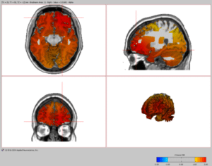
Advanced brain software imagery systems can integrate MRI and EEG data into comprehensive clinical neuroscience scans.
Conditions That Benefit From Combined MRI and EEG Imaging
ADHD and Attention Disorders
ADHD brain imagery often benefits from integrated imaging because attention-related symptoms may involve both:
- Functional brainwave abnormalities
- Structural differences in attention-regulating regions
EEG may reveal:
- Elevated theta activity
- Reduced beta activity
- Functional attention irregularities
MRI may evaluate:
- Structural brain regions associated with executive functioning
- Neurological development patterns
Combining both improves the understanding of cognitive and attention-related disorders.
Patients frequently search for:
- “ADHD brain imaging near me”
- “EEG for ADHD”
- “clinical neuroscience scan Florida”
to learn more about these advanced evaluations.
Epilepsy and Seizure Disorders
EEG is one of the most important tools for detecting seizure activity because it records abnormal electrical discharges in real time.
MRI helps identify:
- Structural lesions
- Brain malformations
- Injury-related abnormalities
Together, MRI and EEG help clinicians:
- Localize seizure origins
- Improve surgical planning
- Guide treatment strategies
Traumatic Brain Injury (TBI)
Concussions and brain injuries may involve:
- Structural damage
- Functional neurological disruptions
MRI can reveal:
- Tissue damage
- Swelling
- Structural abnormalities
EEG may detect:
- Brainwave irregularities
- Functional impairments
- Cognitive processing disruptions
Integrated imaging supports more comprehensive post-injury evaluations.
Cognitive and Memory Disorders
Conditions such as dementia and Alzheimer’s disease often affect:
- Brain structure
- Neural communication pathways
Combining MRI and EEG helps clinicians evaluate:
- Cognitive decline
- Functional connectivity
- Brain atrophy
- Neurological efficiency
Earlier detection may improve long-term treatment planning.
The Role of Neuroscience Software Imagery
Advanced neuroscience software imagery is essential for combining MRI and EEG data effectively.
Modern software systems can:
- Overlay EEG activity onto MRI brain maps
- Create 3D brain imagery
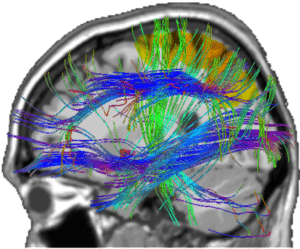
- Analyze neural connectivity
- Detect abnormalities
- Generate detailed clinical reports
These technologies improve:
- Visualization
- Diagnostic interpretation
- Clinical communication
- Personalized treatment planning
At Applied Neuroscience INC., advanced brain software imagery supports comprehensive neurological evaluations and expert clinical reporting.
Benefits of 3D Brain Imagery
Three-dimensional visualization has become an important part of integrated neuroscience imaging.
3D brain imagery helps clinicians:
- Examine brain structures spatially
- Understand functional networks
- Visualize neural communication pathways
- Detect subtle abnormalities
This level of visualization improves both diagnostic analysis and patient understanding.
What Patients Can Expect During MRI and EEG Testing
MRI Procedure
During an MRI:
- Patients lie inside a scanning machine
- Magnetic fields generate detailed brain images
- The procedure is painless and non-invasive
MRI sessions generally last:
- 30–60 minutes
EEG Procedure
During EEG testing:
- Sensors are placed on the scalp
- Brainwave activity is recorded
- Patients may rest quietly or complete tasks
EEG studies typically last:
- 20–90 minutes
Integrated Analysis
Once testing is complete, neuroscience software imagery systems combine the data into detailed clinical reports.
These reports may support:
- Diagnosis
- Treatment planning
- Rehabilitation strategies
- Medical legal evaluations
MRI and EEG in Medical Legal Services
Objective neurological imaging data is increasingly valuable in medical legal cases.
Combined MRI and EEG evaluations may support:
- Brain injury documentation
- Cognitive impairment claims
- Disability assessments
- Neurological expert testimony
- Post-accident evaluations
[Medical Legal Services –https://appliedneuroscienceinc.com/medical-legal-services/]
Comprehensive clinical reports often play a critical role in complex neurological injury cases.
Why Patients in St. Petersburg Are Seeking Integrated Brain Imaging
Patients throughout St. Petersburg and surrounding Florida communities are increasingly searching for:
- “MRI and EEG near me”
- “advanced brain imaging St. Petersburg”
- “clinical neuroscience scan Florida”
- “ADHD brain imagery”
Growing awareness around neurological health, concussion management, and cognitive performance is increasing demand for integrated imaging services.
Frequently Asked Questions
Is MRI or EEG better for ADHD?
Both tests provide different types of information. EEG evaluates functional brain activity, while MRI evaluates brain structure. Together, they may provide more comprehensive insights.
Can EEG detect brain damage?
EEG can detect functional abnormalities in brain activity, but MRI is generally better for identifying structural damage.
Are MRI and EEG safe?
Yes. Both technologies are non-invasive and widely considered safe when performed by qualified professionals.
Why combine MRI and EEG?
Combining MRI and EEG provides both structural and functional information, improving diagnostic accuracy and treatment planning.
How long does integrated imaging take?
The total time depends on the tests performed but may range from 1–3 hours or more.
Why Choose Applied Neuroscience INC.
Applied Neuroscience INC. provides advanced neuroscience imaging services in St. Petersburg, Florida.
Services include:
- MRI and EEG integration
- Clinical neuroscience scans
- ADHD brain imagery
- Brain software imagery
- 3D brain imagery
- Expert clinical reports
- Medical legal services
- Mentoring and consultation
The organization combines advanced imaging technology with exceptional customer service to support comprehensive neurological evaluations.
[Applied Neuroscience INC. –https://appliedneuroscienceinc.com/]
Call to Action
If you are looking for advanced MRI and EEG brain imaging services in St. Petersburg, FL, Applied Neuroscience INC. offers comprehensive neuroscience imaging solutions designed to support accurate neurological evaluations and personalized care.
Contact Applied Neuroscience INC. today to learn more about:
- Clinical neuroscience scans
- MRI and EEG integration
- ADHD brain imaging
- 3D brain imagery
- Advanced neurological diagnostics
Serving St. Petersburg and surrounding Florida communities.