

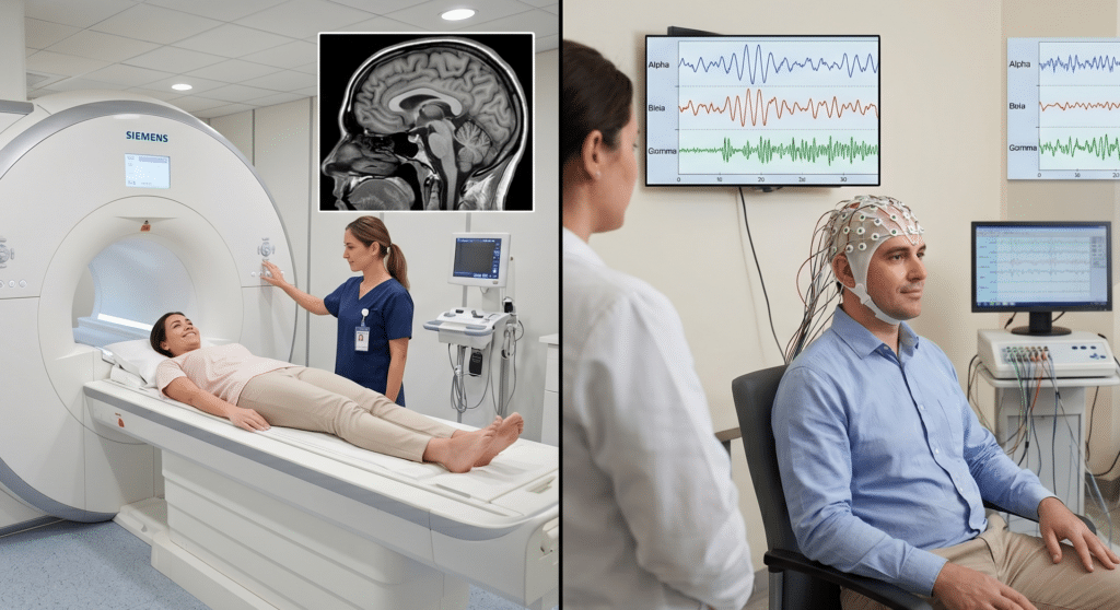
Modern neuroscience is evolving rapidly, and one of the most important advancements in neurological diagnostics is the ability to combine MRI and EEG technologies. By integrating structural brain imaging with real-time electrical activity monitoring, healthcare professionals can gain a more complete understanding of brain function and neurological conditions.
For patients dealing with ADHD, epilepsy, traumatic brain injuries, cognitive disorders, or unexplained neurological symptoms, combining MRI and EEG can provide more accurate diagnoses, better treatment planning, and improved long-term outcomes. At Applied Neuroscience INC. in St. Petersburg, Florida, advanced clinical neuroscience imaging technologies help physicians and specialists see both the structure and function of the brain in ways that traditional standalone imaging cannot achieve.
What Is MRI and EEG Integration?
MRI (Magnetic Resonance Imaging) captures highly detailed images of the brain’s physical structure. It helps identify abnormalities such as:
- Tumors
- Brain injuries
- Structural damage
- Inflammation
- Degenerative conditions
- Stroke-related changes
EEG (Electroencephalogram), on the other hand, measures the brain’s electrical activity in real time. It detects irregular brainwave patterns associated with:
- ADHD
- Epilepsy
- Sleep disorders
- Anxiety
- Cognitive dysfunction
- Neurological abnormalities
When MRI and EEG are combined, physicians gain both anatomical and functional insights into the brain.
This integrated approach allows clinicians to understand not only what the brain looks like, but also how it behaves.
Why Combining MRI and EEG Improves Diagnostic Accuracy
Traditional neurological evaluations often rely on either structural imaging or electrical monitoring separately. However, many neurological and cognitive conditions involve both structural and functional abnormalities.
Combining MRI and EEG helps bridge this gap.
Key Advantages Include:
| MRI Benefits | EEG Benefits |
| Detailed structural imaging | Real-time brain activity monitoring |
| Detects physical abnormalities | Detects functional abnormalities |
| Identifies lesions or damage | Measures brainwave irregularities |
| High-resolution anatomical mapping | Tracks electrical communication |
Together, these technologies create a more comprehensive clinical neuroscience scan.
Conditions That Benefit From Combined MRI and EEG Imaging
ADHD and Attention Disorders
ADHD brain imaging has become increasingly valuable in understanding how different brain regions communicate. While MRI may show structural differences in attention-regulating regions, EEG can reveal irregular brainwave activity patterns commonly associated with ADHD.
This dual analysis helps clinicians:
- Improve diagnostic confidence
- Personalize treatment strategies
- Monitor therapy effectiveness
- Differentiate ADHD from anxiety or learning disorders
Patients searching for advanced ADHD brain imagery near St. Petersburg often benefit from integrated neuroimaging approaches.
Epilepsy and Seizure Disorders
One of the most common uses of combined MRI and EEG technology is epilepsy diagnosis.
MRI identifies structural abnormalities that may trigger seizures, while EEG pinpoints abnormal electrical activity responsible for seizure events.
Together, these scans help:
- Localize seizure origins
- Improve surgical planning
- Reduce diagnostic uncertainty
- Guide medication management
Traumatic Brain Injuries (TBI)
Patients who experience concussions or traumatic brain injuries may continue to suffer symptoms long after a visible injury appears healed.
MRI may reveal subtle structural changes, while EEG detects ongoing disruptions in brain function.
This is particularly important for:
- Sports-related concussions
- Car accident injuries
- Military-related head trauma
- Workplace injuries
Cognitive and Memory Disorders
Neurodegenerative diseases such as Alzheimer’s disease and dementia often affect both brain structure and neural communication pathways.
Combining MRI and EEG allows clinicians to:
- Detect abnormalities earlier
- Track disease progression
- Evaluate cognitive decline more accurately
- Improve long-term treatment planning
How Integrated Brain Imaging Supports Personalized Treatment
One of the greatest advantages of combining MRI and EEG is the ability to personalize care.
Every brain is different. Patients with similar symptoms may show very different imaging results.
Integrated clinical neuroscience scans can help physicians:
- Tailor medication strategies
- Develop individualized therapy plans
- Track treatment progress
- Improve rehabilitation outcomes
- Reduce trial-and-error treatment approaches
For example, ADHD patients may display distinct EEG brainwave patterns that respond differently to behavioral therapies or medication interventions.
This level of precision supports better patient outcomes and more informed medical decisions.
The Role of Advanced Neuroscience Software Imaging
Modern neuroscience software imagery platforms play a critical role in combining MRI and EEG data.
Advanced software can:
- Overlay EEG activity onto MRI brain maps
- Generate 3D brain imagery
- Identify neurological biomarkers
- Analyze functional connectivity
- Visualize abnormal brain activity patterns
These technologies improve diagnostic interpretation and help clinicians explain findings more clearly to patients and families.
At Applied Neuroscience INC., advanced brain software imaging technologies support comprehensive neurological evaluations for physicians, specialists, and medical professionals.
Benefits of 3D Brain Imaging in MRI and EEG Integration
3D brain imagery takes neurodiagnostic analysis to another level.
Instead of reviewing flat scan slices, clinicians can examine:
- Brain regions spatially
- Neural activity distribution
- Functional networks
- Connectivity patterns
This improves:
- Diagnostic clarity
- Surgical planning
- Patient education
- Clinical reporting
Three-dimensional visualization is particularly valuable in complex neurological cases where subtle abnormalities may otherwise go unnoticed.
What Patients Can Expect During the Process
MRI Procedure
MRI scans are non-invasive and painless. Patients lie inside the scanner while detailed brain images are captured using magnetic fields and radio waves.
Typical MRI sessions last:
- 30–60 minutes
EEG Procedure
During EEG testing:
- Small electrodes are placed on the scalp
- Brainwave activity is recorded
- Patients remain awake or perform guided tasks
EEG studies generally take:
- 20–90 minutes, depending on testing requirements
Combined Data Analysis
Once both scans are complete, neuroscience imaging software integrates the information into a comprehensive clinical report.
This report may help physicians:
- Confirm diagnoses
- Identify neurological abnormalities
- Develop treatment strategies
- Support medical legal evaluations
Why Patients in St. Petersburg Are Seeking Advanced Brain Imaging
Healthcare providers and patients throughout St. Petersburg and Pinellas County are increasingly seeking advanced neuroscience imaging services because of growing awareness around:
- ADHD diagnostics
- Cognitive health
- Brain injury assessment
- Functional brain disorders
- Personalized neurological care
Patients often search for:
- “best brain imaging center near me”
- “ADHD brain scan in St. Petersburg”
- “combine MRI and EEG services”
- “clinical neuroscience scan Florida”
Integrated imaging services provide answers that traditional testing methods may miss.
How Combined Imaging Supports Medical Legal Services
Advanced brain imaging is also becoming increasingly important in medical legal evaluations.
Objective MRI and EEG findings may help support:
- Injury documentation
- Cognitive impairment claims
- Disability evaluations
- Neurological expert testimony
- Post-accident assessments
[Medical Legal Services – https://appliedneurosc.wpenginepowered.com/]
These services are especially valuable in complex neurological injury cases requiring detailed clinical documentation.
Frequently Asked Questions
Is combining MRI and EEG safe?
Yes. Both MRI and EEG are non-invasive and widely used diagnostic tools with strong safety records.
Can MRI and EEG diagnose ADHD?
While no single test definitively diagnoses ADHD, combined imaging may provide valuable neurological insights that support comprehensive evaluations.
How long does combined brain imaging take?
Testing time varies depending on the type of evaluation but may range from 1–3 hours total.
Does insurance cover clinical neuroscience scans?
Coverage depends on the patient’s provider, diagnosis, and medical necessity. Patients should verify benefits directly with their insurance carrier.
What makes combined imaging better than traditional scans?
MRI provides structural detail while EEG measures real-time brain activity. Together, they offer a more complete understanding of neurological function.
Why Choose Applied Neuroscience INC. in St. Petersburg, FL
Applied Neuroscience INC. provides advanced clinical neuroscience imaging solutions designed to improve diagnostic accuracy and patient care.
Services include:
- Brain software imagery
- MRI and EEG integration
- Expert clinical reports
- Medical legal services
- Mentoring and consultation
- Advanced neurodiagnostic analysis
The organization’s focus on customer service and advanced neuroscience technology helps physicians and patients access more comprehensive neurological insights.
[Applied Neuroscience INC. –https://appliedneuroscienceinc.com/
Call to Action
If you are looking for advanced brain imaging services in St. Petersburg, FL, Applied Neuroscience INC. offers integrated MRI and EEG diagnostic solutions designed to support accurate neurological evaluations and personalized care.
Contact Applied Neuroscience INC. today to learn more about:
- Clinical neuroscience scans
- ADHD brain imaging
- 3D brain imagery
- Medical legal neurodiagnostic services
- Expert clinical reporting
Serving St. Petersburg and surrounding Florida communities.